Antagonistic muscles – opposing muscles (radial and circular)
Antagonistic muscles are the pair of opposing muscles in the iris that control the size of the pupil. One is a circular muscle layer and the other a radial muscle layer.
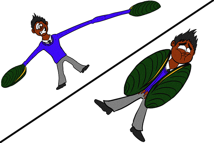
The two mussels (muscles) were antagonistic towards the pupil, first stretching him, then squeezing him.
Radial and circular muscle
Located in the iris, the radial muscle opens the pupil wider, and the circular muscle reduces it, but only one type can work at any one time, while the other relaxes. They get the name “antagonistic” because they oppose each other, working in opposite directions.
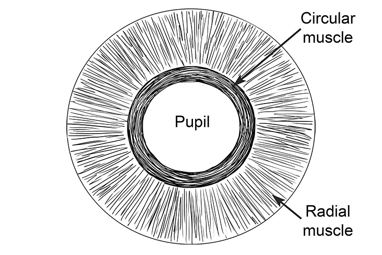
When the circular muscle contracts and the radial muscle relaxes, the pupil reduces in size.
When the radial muscle contracts and the circular muscle relaxes, the pupil opens wider.
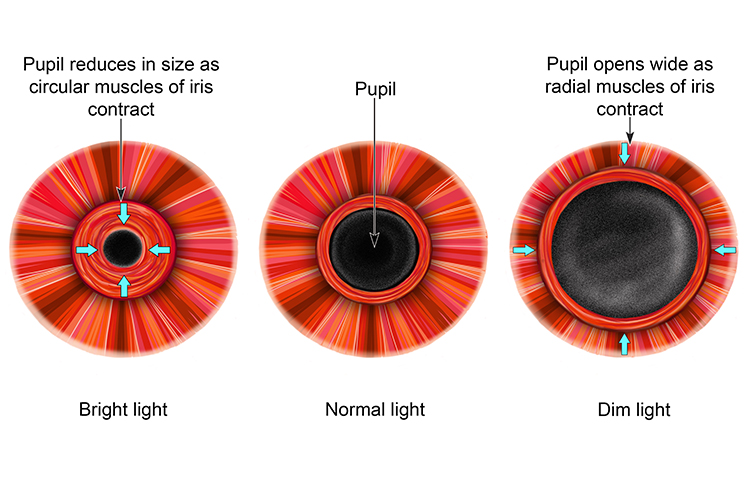
How the antagonistic muscles work to constrict and dilate the pupil.
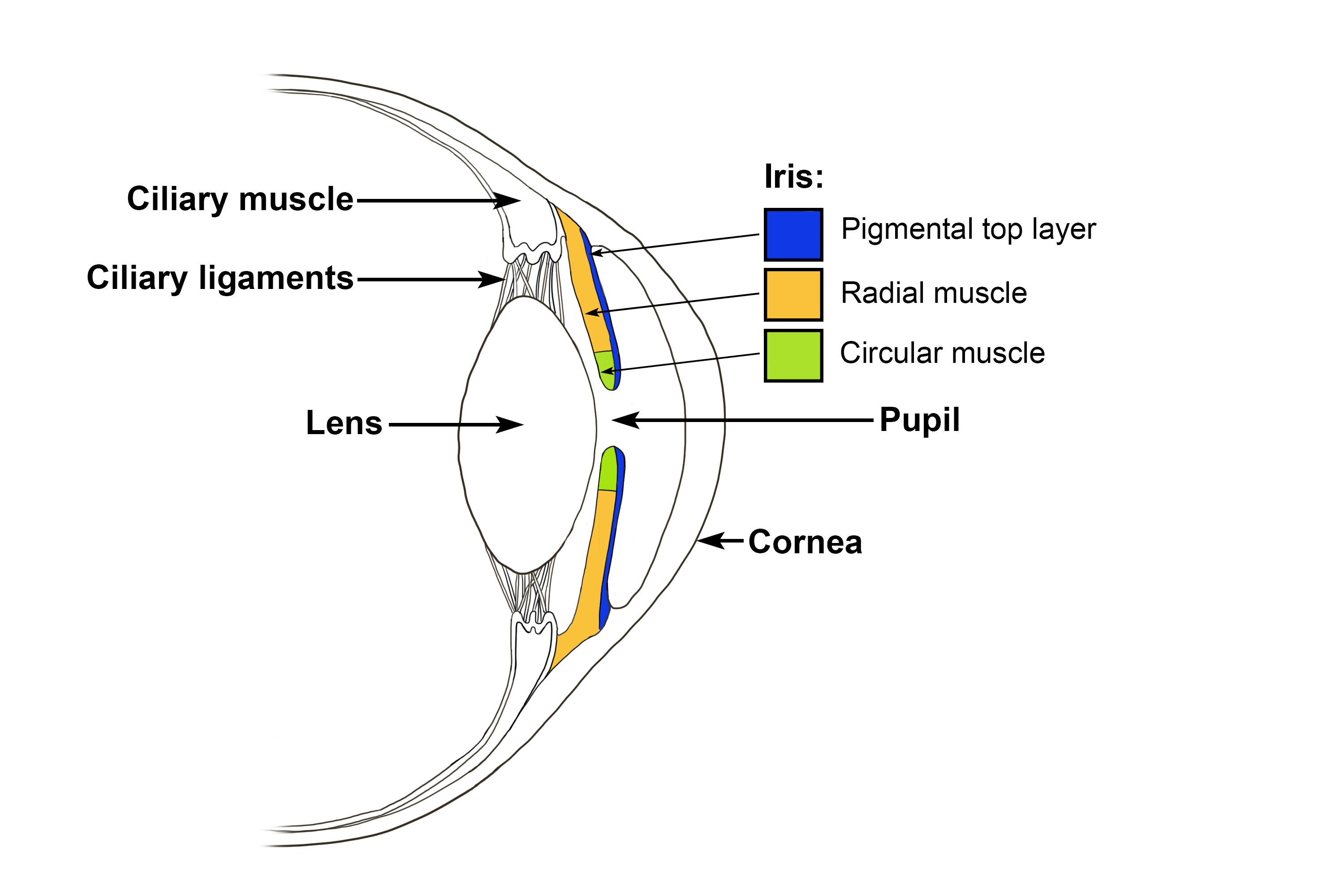
Sectional side view showing position of iris in relation to lens.
NOTE:
These muscles are normally hidden by the pigmented top layer of the iris (which gives your eyes their colour).
